
Secondary curves are concave posteriorly, opposite in direction to the original fetal curvature. Each of these is thus called a primary curve because they are retained from the original fetal curvature of the vertebral column.Ī secondary curve develops gradually after birth as the child learns to sit upright, stand, and walk. In the adult, this fetal curvature is retained in two regions of the vertebral column as the thoracic curve, which involves the thoracic vertebrae, and the sacrococcygeal curve, formed by the sacrum and coccyx. Primary curves are retained from the original fetal curvature, while secondary curvatures develop after birth.ĭuring fetal development, the body is flexed anteriorly into the fetal position, giving the entire vertebral column a single curvature that is concave anteriorly. The four adult curvatures are classified as either primary or secondary curvatures. They then spring back when the weight is removed. When the load on the spine is increased, by carrying a heavy backpack for example, the curvatures increase in depth (become more curved) to accommodate the extra weight. These curves increase the vertebral column’s strength, flexibility, and ability to absorb shock. The adult vertebral column does not form a straight line, but instead has four curvatures along its length (see Figure 1). In a full-grown giraffe, each cervical vertebra is 11 inches tall. This means that there are large variations in the size of cervical vertebrae, ranging from the very small cervical vertebrae of a shrew to the greatly elongated vertebrae in the neck of a giraffe. However, the sacral and coccygeal fusions do not start until age 20 and are not completed until middle age.Īn interesting anatomical fact is that almost all mammals have seven cervical vertebrae, regardless of body size. Similarly, the coccyx, or tailbone, results from the fusion of four small coccygeal vertebrae.

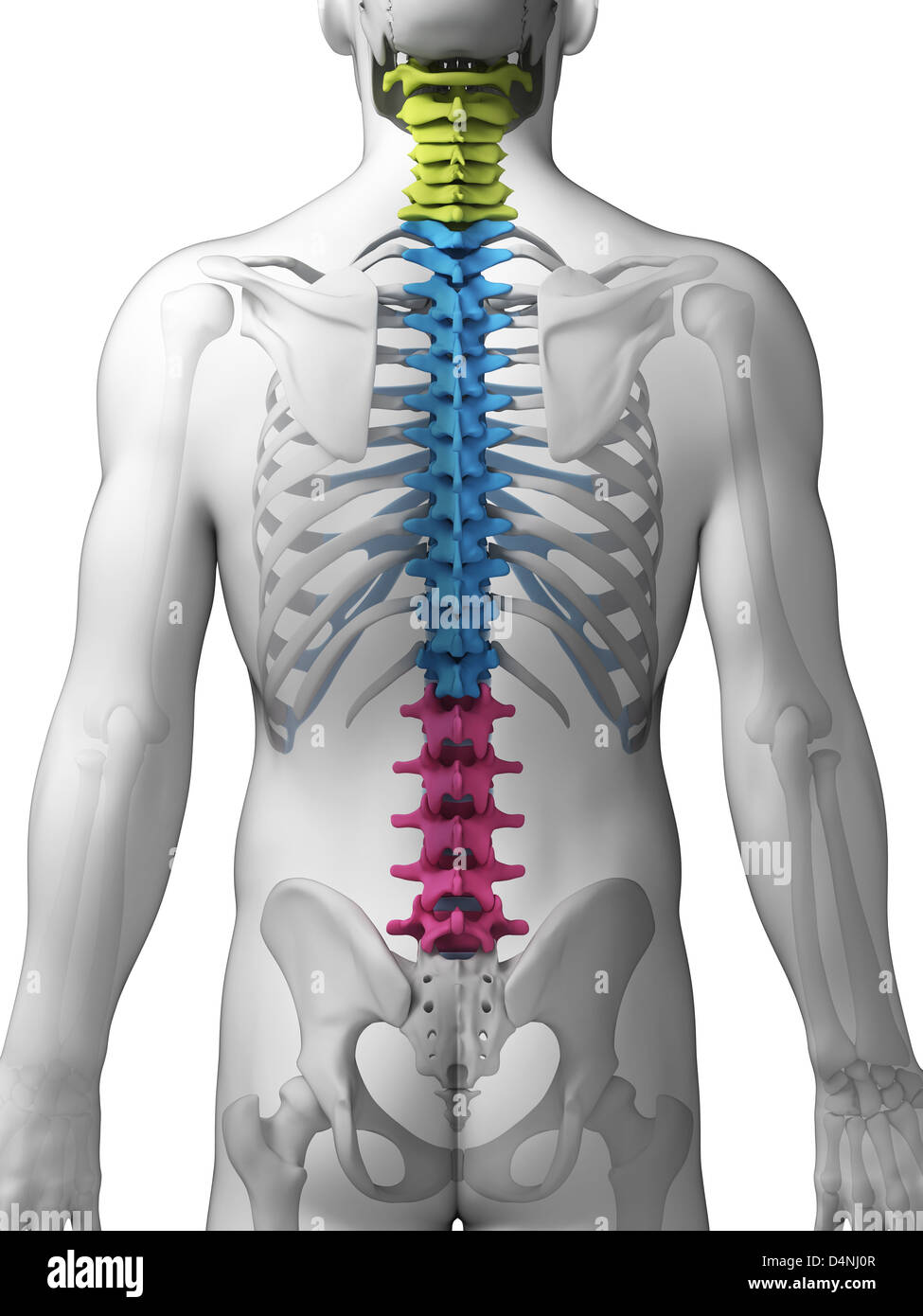
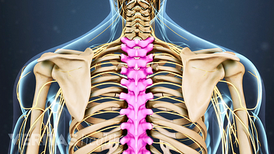
The single sacrum, which is also part of the pelvis, is formed by the fusion of five sacral vertebrae. The lower back contains the L1–L5 lumbar vertebrae. Below these are the 12 thoracic vertebrae, designated T1–T12. Inferiorly, C1 articulates with the C2 vertebra, and so on.

Superiorly, the C1 vertebra articulates (forms a joint) with the occipital condyles of the skull. In the neck, there are seven cervical vertebrae, each designated with the letter “C” followed by its number. The vertebral column is subdivided into five regions, with the vertebrae in each area named for that region and numbered in descending order.
Sections of the spine plus#
The vertebral column originally develops as a series of 33 vertebrae, but this number is eventually reduced to 24 vertebrae, plus the sacrum and coccyx. The vertebral column is curved, with two primary curvatures (thoracic and sacrococcygeal curves) and two secondary curvatures (cervical and lumbar curves). The vertebrae are divided into three regions: cervical C1–C7 vertebrae, thoracic T1–T12 vertebrae, and lumbar L1–L5 vertebrae. The adult vertebral column consists of 24 vertebrae, plus the sacrum and coccyx.


 0 kommentar(er)
